Diagnosis & Treatments

How is Bile Duct Cancer diagnosed?

Diagnosis of Bile Duct Cancer
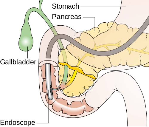
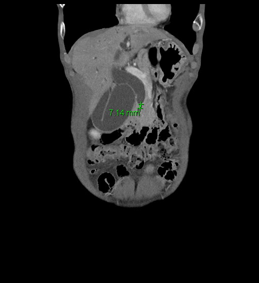
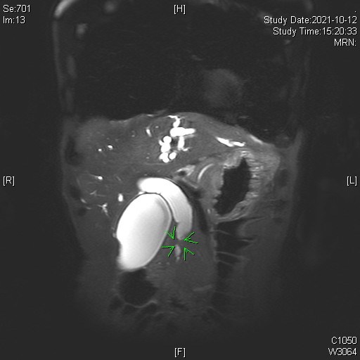
Bile duct cancer(1).pngBile duct cancer(2).jpgBile duct cancer(3).jpg


1. Multiphase CT and MRI

  • High-resolution contrast-enhanced CT and MRI scans are first-line tools for evaluating biliary masses, ductal obstruction, vascular involvement, and metastatic spread.
  • MRI with MRCP (Magnetic Resonance Cholangiopancreatography) is particularly useful for visualizing the biliary tree non-invasively and assessing the level and extent of bile duct strictures or masses.

2. Endoscopic Ultrasound (EUS)

  • EUS provides high-resolution imaging of the distal bile duct and ampullary region. It is especially useful for detecting small or infiltrative tumors and can guide tissue acquisition via fine-needle aspiration (FNA).

3. Histological Tissue Biopsy (EUS-FNA/B, ERCP sampling, PTC biopsy)

  • A definitive diagnosis relies on pathology. Tissue samples can be obtained via EUS-guided biopsy, brushings or forceps biopsy during ERCP, or percutaneous approaches such as PTC-guided biopsy.

4. Endoscopic Retrograde Cholangiopancreatography (ERCP)

  • ERCP is used for both diagnostic and therapeutic purposes, allowing ductal visualization, cytologic sampling, and stent placement for biliary obstruction.
  • In select cases, peroral digital cholangioscopy enables real-time visualization and targeted biopsies, improving diagnostic yield for biliary strictures and intraductal lesions.

5. Blood Samples

  • Tumor markers such as CA 19-9 and CEA may support diagnosis but have limited specificity.
  • Liquid biopsy techniques, including circulating tumor DNA (ctDNA) and exosome analysis, are emerging tools for non-invasive tumor profiling.

Stages of Bile Duct Cancer

Staging of biliary tract cancer depends on the anatomical subtype: intrahepatic cholangiocarcinoma, perihilar cholangiocarcinoma, distal cholangiocarcinoma, gallbladder cancer, or ampullary cancer.


The TNM classification considers:

  • T (Tumor size and extent of invasion)
  • N (Regional lymph node involvement)
  • M (Distant metastasis)


Each subtype has distinct staging criteria under AJCC 8th edition. Imaging (CT, MRI, PET), diagnostic laparoscopy, and pathology are used to determine clinical stage and resectability.

Diagnosis & Treatments

How is Bile Duct Cancer treated?

Treatments for Bile Duct Cancer

Multidisciplinary Approach

Our hospital adopts a multidisciplinary care model involving specialists from gastroenterology, surgery, medical oncology, radiation oncology, radiology, and pathology. For patients requiring coordinated decision-making—such as those with complex, borderline resectable, or advanced disease—cases are reviewed in a Pancreatobiliary Cancer Center-led Multidisciplinary Meeting, where experts from each department collaborate to establish the most appropriate and individualized treatment plan based on current clinical guidelines and patient-specific factors.


Surgery

Surgical resection offers the best chance for long-term survival in localized disease.

  • Intrahepatic cholangiocarcinoma: Liver resection with regional lymphadenectomy
  • Perihilar cholangiocarcinoma: Extended hepatectomy with bile duct resection and hepaticojejunostomy
  • Distal cholangiocarcinoma / Ampullary cancer: Pancreaticoduodenectomy (Whipple procedure)
  • Gallbladder cancer: Cholecystectomy with liver wedge resection and lymphadenectomy if indicated
  • Vascular reconstruction may be required in select cases with vessel involvement.


※ Adjuvant therapy is typically recommended postoperatively to reduce recurrence.


Chemotherapy

Chemotherapy is a key component in both adjuvant and advanced disease settings.

  • Adjuvant chemotherapy:Capecitabine is commonly used following curative resection.
  • Neoadjuvant chemotherapy: It is not yet considered standard of care but may be selectively applied, particularly in borderline resectable or locally advanced disease. 
  • Systemic chemotherapy for unresectable/metastatic disease:


Common regimens include:


Primary treatment

  • Gemcitabine + cisplatin + durvalumab
  • Gemcitabine + cisplatin + pembrolizumab
  • Gemcitabine + cisplatin


Subsequent-line therapy

  • FOLFOX
  • Liposomal irinotecan + fluorouracil + leucovorin
  • Nivolumab
  • etc.


Targeted Therapy and Immunotherapy

Targeted and immune-based therapies are increasingly being explored in biliary tract cancer (BTC), especially in patients with advanced or unresectable disease. Treatment options are guided by molecular profiling.


Commonly used or emerging targeted agents include:


FGFR2 fusions or rearrangements

  • Treated with pemigatinib, futibatinib, or other FGFR inhibitors (especially in intrahepatic cholangiocarcinoma)


IDH1 mutations

  • Targeted by ivosidenib


HER2 overexpression or amplification

  • May be treated with HER2-targeted therapies such as trastuzumab and pertuzumab, often in combination with chemotherapy


MSI-H or dMMR tumors

  • May benefit from immune checkpoint inhibitors such as pembrolizumab


Comprehensive molecular profiling (e.g., next-generation sequencing) is recommended for patients with advanced BTC to identify potential targets. Additional investigational therapies are available through clinical trials, including agents targeting BRAF, NTRK, PIK3CA, and others.


Supportive and Palliative Care


Supportive care is critical to improving patient comfort and quality of life:

  • Biliary drainage: via ERCP or percutaneous transhepatic biliary drainage (PTBD)
  • Pain control and symptom management
  • Nutritional support and psychosocial services