Diagnosis & Treatments

How is Pancreatic Cancer diagnosed?

Diagnosis of Pancreatic Cancer
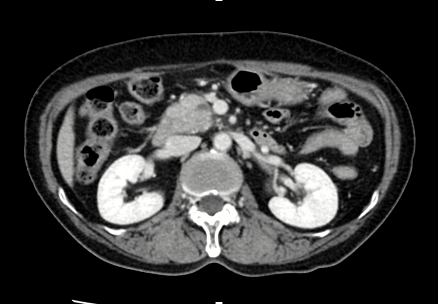
Pancreatic cancer(1).png

CT

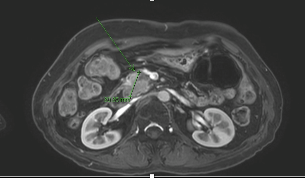
Pancreatic cancer(2).png

MRI

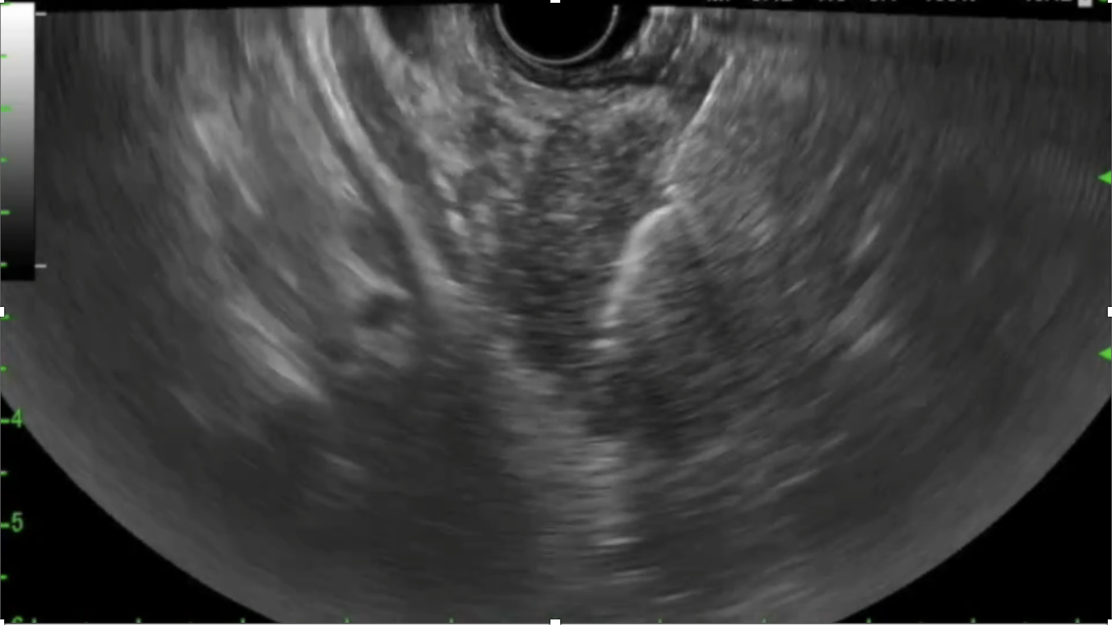
Pancreatic cancer(3).png

EUS-guided biopsy

1. Multiphase CT and MRI

  • These are standard first-line imaging techniques that provide high-resolution visualization of pancreatic masses, local invasion, and metastatic spread.
  • Multiphase CT (pancreatic protocol) remains the backbone for tumor staging and surgical planning.
  • MRI, particularly with MRCP and diffusion-weighted imaging, offers superior soft tissue contrast and is especially helpful in detecting is attenuating tumors. It also enables non-invasive visualization of the pancreatic duct and bile duct, making it highly valuable in evaluating ductal obstruction. Furthermore, MRI is preferred in patients with renal impairment or iodine contrast allergy and provides multiparametric data that can aid in tumor characterization and staging.

2. Endoscopic Ultrasound (EUS) 

  • Endoscopic ultrasound (EUS) provides high-resolution, real-time imaging of the pancreas and surrounding structures. It is particularly valuable for detecting small or deeply located pancreatic lesions and guiding further diagnostic procedures.

3. Histological Tissue Biopsy (EUS-FNA/B, ERCP sampling)

  • Definitive diagnosis remains reliant on pathology examination of tissue samples. EUS guided fine-needle aspiration and biopsies or ERCP-based ductal brushings and biopsies are commonly used. 

4. Blood samples

  • Traditional tumor markers like CA?19-9 and CEA are included in the diagnostic work-up but have limitations in sensitivity and specificity.
  • Liquid biopsy techniques like circulating tumor DNA (ctDNA) and exosome analysis can detect molecular fingerprints of tumors with minimal invasiveness, and are being evaluated for high-risk screening.


Stages of Pancreatic Cancer

Pancreatic Cancer Staging (AJCC 8th Edition)

Pancreatic cancer staging helps determine the extent of disease and guides treatment planning. The American Joint Committee on Cancer (AJCC) 8th edition defines stages based on tumor size (T), lymph node involvement (N), and distant metastasis (M).


Stage 0 – Carcinoma in Situ (Tis; N0; M0)

Abnormal (pre-cancerous) cells are found in the lining of the pancreatic ducts. These cells have not invaded deeper tissues or spread. This stage is also called carcinoma in situ and may develop into invasive cancer over time.


Stage I – Localized Disease (T1–T2; N0; M0)

Cancer is confined to the pancreas and has not spread to lymph nodes or distant sites.

  • Stage IA: The tumor is 2 cm or smaller (T1).
  • Stage IB: The tumor is larger than 2 cm but not more than 4 cm (T2).

Stage II – Locally Advanced without Distant Spread

The tumor may be larger and/or may have spread to nearby lymph nodes, but not to distant organs.

  • Stage IIA: Tumor is >4 cm but has not spread to lymph nodes (T3; N0; M0).
  • Stage IIB: Tumor of any size (T1–T3) has spread to 1–3 regional lymph nodes (N1), but not to distant sites.

Stage III – Locally Advanced with Extensive Nodal or Vascular Involvement

The cancer involves major arteries near the pancreas (e.g., celiac axis or superior mesenteric artery), or has spread to 4 or more regional lymph nodes.

  • Includes T4 tumors (involving major vessels) or any tumor with N2 nodal status (≥4 regional lymph nodes), but no distant metastasis (M0).

Stage IV – Metastatic Disease (Any T; Any N; M1)

The cancer has spread to distant sites such as the liver, lungs, or peritoneal cavity. Tumor size and lymph node status may vary, but the presence of distant metastasis defines this stage.

Diagnosis & Treatments

How is Pancreatic Cancer treated?

Treatments for Pancreatic Cancer

1. Surgery

Surgery is the only potentially curative treatment for pancreatic cancer. It is usually considered in patients with localized (resectable) or borderline resectable disease.


Whipple procedure (Pancreaticoduodenectomy)

Performed when the tumor is located in the head of the pancreas. It involves removal of the pancreatic head, part of the small intestine, gallbladder, and bile duct.


Distal Pancreatectomy

Used for tumors in the body or tail of the pancreas. The spleen is often removed as well.


Total Pancreatectomy

Removal of the entire pancreas, usually performed in select cases.


Vascular Resection and Reconstruction

In borderline resectable cases, involved blood vessels (e.g., portal vein or superior mesenteric vein) may be resected and reconstructed during surgery.


*Surgery is typically followed by adjuvant therapy to reduce the risk of recurrence.


2. Chemotherapy

Chemotherapy plays a central role in the treatment of pancreatic cancer and may be used in various settings:


Adjuvant chemotherapy

Given after surgery to eliminate microscopic residual disease. Common regimens include mFOLFIRINOX or gemcitabine + capecitabine.


Neoadjuvant chemotherapy

Used before surgery, especially in borderline resectable cases, to shrink the tumor and improve surgical outcomes. Common regimens include FOLFIRINOX.


Systemic chemotherapy for unresectable/metastatic disease

In advanced-stage cancers, chemotherapy is the primary treatment. Common regimens include:


  • FOLFIRINOX (oxaliplatin, irinotecan, leucovorin, and 5-FU)
  • Gemcitabine + nab-paclitaxel
  • NALIRIFOX (liposomal irinotecan, 5-FU, leucovorin, and oxaliplatin)
  • Gemcitabine alone (for frail or elderly patients)
  • Liposomal irinotecan + 5-FU + leucovorin (If prior gemcitabine therapy)


3. Targeted Therapy and Immunotherapy

PARP inhibitors (e.g., olaparib)

Indicated in selected patients with BRCA1/2 mutations after response to platinum-based chemotherapy.


Immune checkpoint inhibitors

For patients with microsatellite instability-high (MSI-H) or mismatch repair-deficient (dMMR) tumors, though these are rare in pancreatic cancer.


Clinical Trials

Patients may benefit from participation in ongoing clinical trials evaluating novel agents.


4. Radiation Therapy

Radiation therapy may be used in selected patients to control local tumor growth, relieve symptoms, or as part of a combined treatment approach.


Proton Therapy (Proton Beam Radiation)

An advanced form of radiation that uses protons instead of X-rays. Proton therapy delivers radiation with remarkable precision, reducing radiation exposure to nearby healthy organs such as the liver, kidneys, and intestines.

It is particularly beneficial for locally advanced tumors located near critical structures, and may reduce treatment-related side effects in selected patients.


Stereotactic Body Radiation Therapy (SBRT)

Delivers high-dose radiation in a few fractions with precision targeting.


Conventional chemoradiation

Often used with radiosensitizing chemotherapy (e.g., 5-FU or capecitabine) in locally advanced or borderline resectable disease.


5. Supportive and Palliative Care

Comprehensive supportive care is essential to improve quality of life:


  • Pain management
  • Nutritional support 
  • Management of biliary or gastric obstruction (e.g., stenting via ERCP or EUS-guided procedures)
  • Psychosocial and palliative care services


6. Multidisciplinary Approach

Our hospital adopts a multidisciplinary care model involving specialists from gastroenterology, surgery, medical oncology, radiation oncology, radiology, and pathology. For patients requiring coordinated decision-making—such as those with complex, borderline resectable, or advanced disease—cases are reviewed in a Pancreatobiliary Cancer Center-led Multidisciplinary Meeting, where experts from each department collaborate to establish the most appropriate and individualized treatment plan based on current clinical guidelines and patient-specific factors.